Difference between revisions of "HFE"
(→Protein Sequence) |
|||
| Line 25: | Line 25: | ||
[[File:HFE-beta2M-2.png|300px]] | [[File:HFE-beta2M-2.png|300px]] | ||
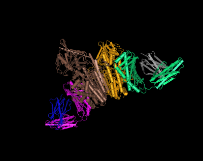
| − | HFE (magenta and green) tertiary complex with beta2M (blue and gray) and TfR (yellow and brown) | + | HFE (magenta and green) tertiary complex with beta2M (blue and gray) and TfR (yellow and brown)<ref>Bennett, M. J., Lebrón, J. A., & Bjorkman, P. J. (2000). Crystal structure of the hereditary haemochromatosis protein HFE complexed with transferrin receptor. Nature, 403(6765), 46-53.</ref> |
[[File:HFE-beta2M-TfR-2.png|400px]] | [[File:HFE-beta2M-TfR-2.png|400px]] | ||
| Line 32: | Line 32: | ||
[[File:HFE protter.png|400px]] | [[File:HFE protter.png|400px]] | ||
| + | |||
| + | <references/> | ||
Revision as of 10:09, 18 July 2014
HFE is named after High Fe (iron). The gene products known function is to regulate iron absorption by interacting with the transferrin receptor (TfR). When the normal function is disrupted by mutation the result can be HFE hereditary hemochromatosis or iron overload.
DNA Sequence
HFE is located on chromosome 6 in humans.
RNA Sequence
There are several alternatively spliced variants of HFE.
Protein Sequence
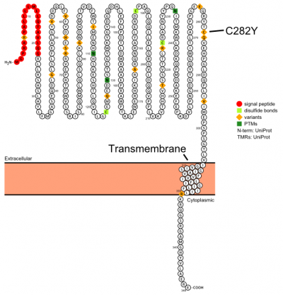
The protein has a signal sequence and transmembrane domain. It forms a tertiary complex with beta2-microglobulin (beta2M) and TfR. HFE has sequence and structural similarity to MHC class I-type proteins.
NCBI Protein[1]
>gi|4504377|ref|NP_000401.1| hereditary hemochromatosis protein isoform 1 precursor [Homo sapiens]
MGPRARPALLLLMLLQTAVLQGRLLRSHSLHYLFMGASEQDLGLSLFEALGYVDDQLFVFYDHESRRVEP
RTPWVSSRISSQMWLQLSQSLKGWDHMFTVDFWTIMENHNHSKESHTLQVILGCEMQEDNSTEGYWKYGY
DGQDHLEFCPDTLDWRAAEPRAWPTKLEWERHKIRARQNRAYLERDCPAQLQQLLELGRGVLDQQVPPLV
KVTHHVTSSVTTLRCRALNYYPQNITMKWLKDKQPMDAKEFEPKDVLPNGDGTYQGWITLAVPPGEEQRY
TCQVEHPGLDQPLIVIWEPSPSGTLVIGVISGIAVFVVILFIGILFIILRKRQGSRGAMGHYVLAERE
NCBI Structure[2]
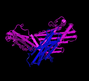
HFE (magenta) tertiary complex with beta2M (blue)
HFE (magenta and green) tertiary complex with beta2M (blue and gray) and TfR (yellow and brown)[1]
Plot in Protter[3] with UniProt accession: Q30201
- ↑ Bennett, M. J., Lebrón, J. A., & Bjorkman, P. J. (2000). Crystal structure of the hereditary haemochromatosis protein HFE complexed with transferrin receptor. Nature, 403(6765), 46-53.


