Agar Diffusion
A simple demonstration of diffusion through agar media at room temperature to visually illustrate that viruses diffuse through plate media. This sets up background knowledge that can be important later. Most plaques only form in a limited window of time when plates are set up and the bacteria are actively dividing. Once this phase has past and the bacteria enter stationary phase the plaques stop growing in size but the virus particles still diffuse through the media. Picking a plaque from an older plate may contain a mix of different kinds of virus particles, especially from nearby plaques (which is why it is important for a plaque to be well isolated from the others).
Red Food Coloring
This was done with a single drop of food coloring containing FD&C Red 3 and Red 40. Be careful not to disturb the plate and cause the drop to run until it is completely absorbed into the media.
After sitting overnight the liquid was completely absorbed into the media and the plate can be inverted.
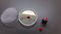
Red Food Coloring, Starting Drop
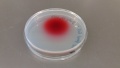
Red Food Coloring, one day later, inverted

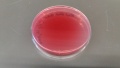
Red Food Coloring, two days later, inverted
Red Food Coloring, one week later, inverted


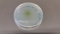
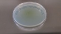
Loading Dye
This was done with 5 ul of gel loading dye (normally used for electrophoresis). Be careful not to disturb the plate and cause the drop to run until it is completely absorbed into the media. The loading dye quickly became less clear than the food coloring.
After sitting overnight the liquid was completely absorbed into the media and the plate can be inverted. After one week the dye was too faint to be visible.
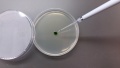
Loading Dye, Starting Drop
Loading Dye, One day later, inverted
Loading Dye, Two days later, inverted


Notes
Carbon black has a particle size closer to that of a virus and might be more appropriate to model viral particle diffusion. There are also a lot of other factors that are not controlled here such as the charges of the particles and interactions with the media as well as the effects of having bacteria in the media.
What Links Here
- SEA-PHAGES (← links)