Michael Wallstrom and Áki Láruson made some scanning electron microscope images of a new species of marine sponge we are working on (it is associated with invasive algal mats here in Hawai'i). I can't resist sharing a few of them here but I am saving the best for the publication we are working on.
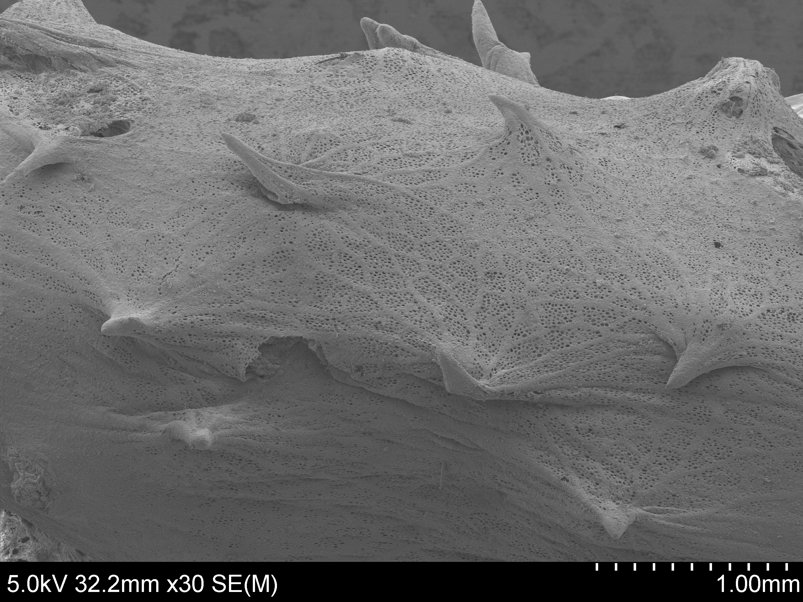
Above is the surface of the sponge. If you look closely you can see the tiny ostia pores in the surface.
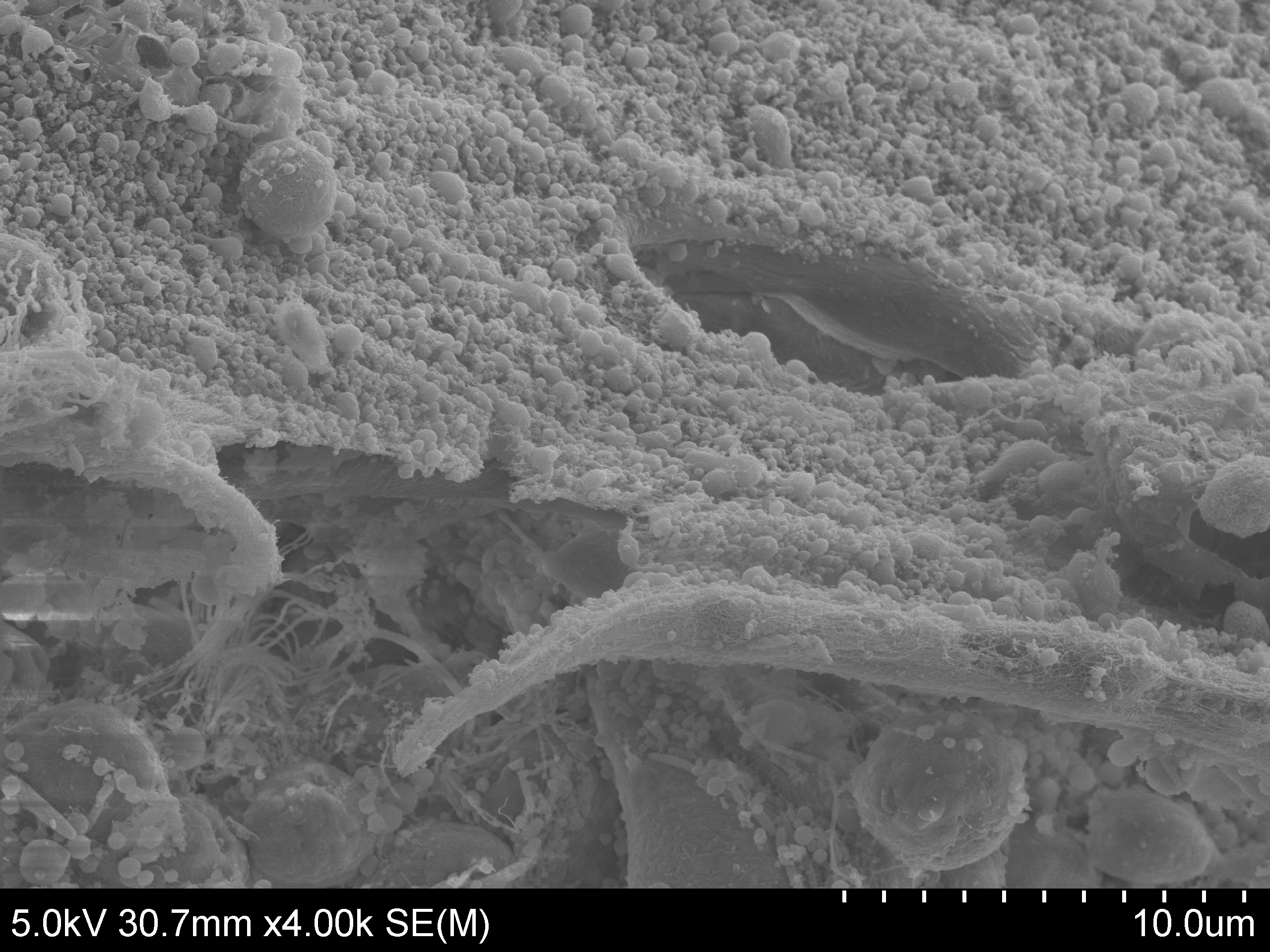
A closeup on the ostia, one is in cross section to the interior of the sponge.
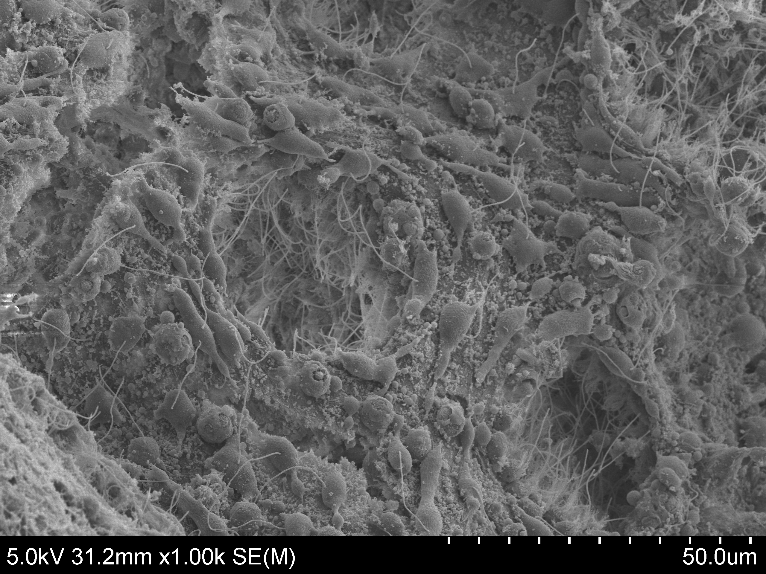
Above, you can see two types of cells inside the sponge. The choanocytes use flagella (the threads) to move water through the sponge and filter food particles out of the seawater; amoebocytes crawl around and transport nutrients to other cells (among other functions).
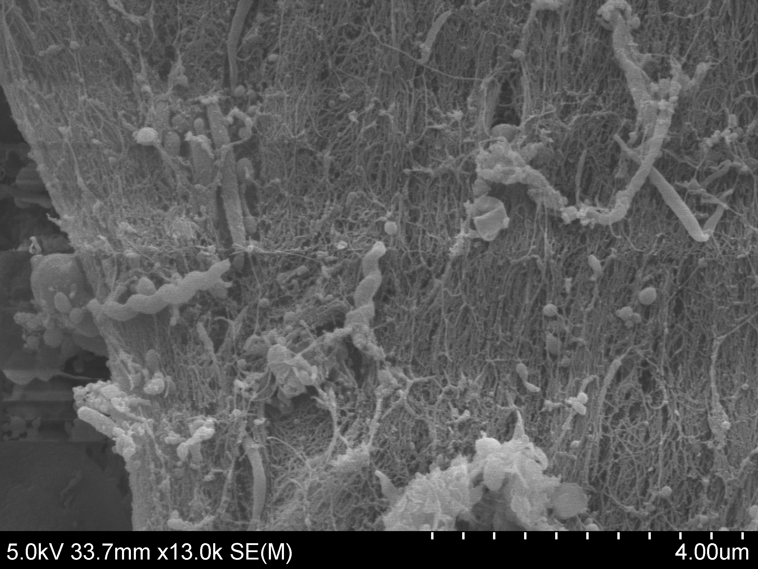
In the above image, at the highest magnification for these images, you can see bacteria that are living in the sponge. The spiral objects are spirochaetes; some of these cause diseases in humans like Lyme disease, syphilis, relapsing fever, and leptospirosis.